Tag: Medical Imaging

  • How AI-Powered Diagnostic Tools Are Revolutionizing Real-Time Healthcare

    How AI-Powered Diagnostic Tools Are Revolutionizing Real-Time Healthcare

    Artificial intelligence is rapidly transforming the way medical professionals diagnose and treat patients. By analyzing medical scans, symptoms, and patient records within seconds, AI-powered diagnostic tools now support faster and more accurate clinical decisions. These systems do not replace human clinicians but act as powerful assistants, enabling doctors to focus on complex cases while reducing repetitive workloads.

    AI Reads Medical Scans Faster

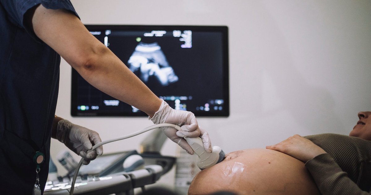
    One of the most impactful applications of AI in healthcare is the rapid analysis of medical imaging. From X-rays and CT scans to MRIs and ultrasounds, AI algorithms can detect tumors, fractures, strokes, lung diseases, and infections. The technology quickly flags abnormalities, allowing radiologists and specialists to prioritize urgent cases and reduce diagnostic delays.

    Continuous Patient Monitoring

    AI systems now monitor patients in real time, especially in intensive care units and through wearable devices. By continuously analyzing vital signs such as heart rate, oxygen levels, blood pressure, and glucose levels, AI can alert healthcare providers to sudden health risks, enabling early intervention and potentially saving lives.

    Smarter Diagnosis Through Data Integration

    Modern AI diagnostic platforms combine multiple data sources—medical images, electronic health records, lab reports, ECGs, and patient-reported symptoms. By cross-referencing these inputs, AI can suggest possible diagnoses and prioritize critical conditions, helping doctors make more informed decisions faster.

    Key Areas Where AI Excels

    Hospitals are increasingly deploying AI in specialties such as cardiology, oncology, radiology, neurology, ophthalmology, and emergency care. In these fields, AI improves early disease detection, speeds up treatment planning, and enhances overall clinical workflow.

    Benefits for Doctors and Patients

    The integration of AI reduces diagnostic errors, streamlines triage, and automates routine data review. This allows doctors to dedicate more time to direct patient care, improving both efficiency and patient satisfaction.

    Important Limitations to Consider

    Despite its promise, AI still has significant limitations. It can miss clinical context, reflect biases present in training data, or produce incorrect recommendations. Therefore, physicians must always verify AI-generated suggestions before making treatment decisions. Responsible implementation requires continuous oversight and validation.

    India Leading the Way in AI Healthcare

    In India, hospitals in cities like Hyderabad and Bengaluru are actively testing AI systems in cardiology, oncology, and emergency medicine. These initiatives aim to improve diagnosis speed and patient outcomes, positioning the country as a hub for AI-driven healthcare innovation.

    The Future: AI as a Clinical Assistant

    Experts agree that the future of medicine lies in collaboration between AI and human expertise. AI works best as a clinical assistant—enhancing decision-making, boosting efficiency, and supporting doctors rather than replacing their irreplaceable judgment and empathy.

  • PLATON AI Camera Reconstructs Particle Paths Using Just Five Photons

    PLATON AI Camera Reconstructs Particle Paths Using Just Five Photons

    Scientists at ETH Zurich and EPFL have developed an AI-powered camera system that can reconstruct the paths of subatomic particles using only a handful of photons. The technology, called PLATON, could simplify particle detectors by replacing millions of conventional detector elements with a single light-emitting block and artificial intelligence. The findings were published in Nature Communications.

    How PLATON Works

    Particle detectors help scientists study elementary particles by tracking how they move through matter. Most existing detectors use scintillators, materials that emit tiny flashes of light when struck by charged particles. To pinpoint where these interactions take place, the scintillator is divided into millions of small segments connected to optical fibres and photon sensors. While this design offers high precision, it also increases the complexity and cost of large-scale detectors. Experiments such as Japan’s T2K neutrino project and CERN’s LHCb rely on these segmented detector systems to accurately track particle interactions.

    PLATON removes the need for millions of detector elements. Instead, it uses a single, unsegmented scintillator block and reconstructs the particle’s trajectory by analysing the emitted light.

    Inside the PLATON System

    The system combines a plenoptic, or light-field, camera with a single-photon avalanche diode (SPAD) sensor that detects individual photons generated inside the scintillator. Unlike conventional cameras, a light-field camera records both the intensity of light and the direction from which it arrives. The research team built the prototype by mounting a micro-lens array directly on the SwissSPAD2 imaging sensor. The setup captures extremely faint light signals while filtering background noise through gated photon detection.

    During laboratory tests, the prototype reconstructed particle interactions using as few as five detected photons, with the experimental results closely matching computer simulations.

    Potential Applications Beyond Physics

    The researchers also developed a transformer-based neural network to analyse where and when photons are detected. Instead of processing text, the AI identifies patterns in photon data to reconstruct the original particle path in three dimensions. Simulations suggest a future version of PLATON could achieve sub-millimetre spatial resolution inside a 10×10×10 cm detector. A larger one-cubic-metre detector is expected to deliver resolution of only a few millimetres, comparable to today’s advanced scintillator detectors despite using far fewer components.

    The researchers also see potential beyond particle physics. They have filed three patents covering the use of PLATON in positron emission tomography (PET) scanners. The technology could improve medical imaging by reconstructing faint light signals more accurately while reducing the complexity of detector systems.

  • How AI Tools Are Revolutionizing Healthcare Business Operations

    How AI Tools Are Revolutionizing Healthcare Business Operations

    Artificial intelligence is rapidly reshaping the healthcare industry, offering powerful tools that improve efficiency, accuracy, and patient outcomes. From automating clinical documentation to accelerating drug discovery, AI is helping healthcare providers deliver better care while reducing costs and administrative burdens. Below are the key areas where AI is making a transformative impact.

    Clinical Documentation Automation

    AI tools automate medical note-taking, reducing paperwork and improving documentation accuracy. This streamlines clinical workflows and allows physicians to spend more time with patients.

    Medical Imaging Analysis

    AI-powered platforms detect abnormalities in X-rays, CT scans, and MRIs faster than traditional methods, enabling earlier diagnoses and significantly improving radiology efficiency.

    Predictive Healthcare Analytics

    Advanced AI analyzes patient records and historical data to predict diseases, optimize treatments, improve resource allocation, and enhance clinical decision-making.

    Precision Medicine

    AI combines genomic, clinical, and molecular data to recommend personalized treatments, helping healthcare providers deliver targeted therapies with better patient outcomes.

    Digital Pathology Support

    AI-assisted pathology tools analyze tissue samples accurately, improve diagnostic consistency, accelerate disease detection, and support pharmaceutical research.

    Administrative Workflow Automation

    AI streamlines appointment scheduling, billing, insurance processing, and patient record management, reducing operational costs while improving organizational productivity.

    Patient Engagement Solutions

    AI-powered virtual assistants provide instant responses, appointment reminders, medication guidance, and personalized health information, improving patient satisfaction and communication.

    Drug Discovery Acceleration

    AI rapidly analyzes biological data, identifies potential drug candidates, shortens research timelines, reduces development costs, and speeds pharmaceutical innovation.

    Emergency Care Optimization

    AI detects critical conditions in real time, alerts specialists immediately, prioritizes urgent cases, and improves treatment speed for life-threatening medical emergencies.

  • MIT’s Latest Breakthroughs in 3D Technology: Augmented Reality, Bioprinting, and Holography

    MIT’s Latest Breakthroughs in 3D Technology: Augmented Reality, Bioprinting, and Holography

    MIT continues to push the boundaries of three-dimensional technology across multiple disciplines. From medical imaging to advanced manufacturing and computer vision, researchers at the institute are developing solutions that transform how we interact with the physical and digital worlds. Below is a curated overview of recent innovations in 3D technology from MIT labs.

    Medical and Health Applications

    An augmented reality system developed at MIT makes medical ultrasounds easier to interpret by generating a real-time 3D representation of the imaged object. Another project leverages machine learning to model fetal shape and movements in 3D, aiding doctors in detecting abnormalities. In bioprinting, the MagMix magnetic mixer enables scalable 3D printing of tissues, and a deep-learning model predicts cellular formation in fruit flies, potentially applicable to human tissues.

    Photonics and Manufacturing

    MIT engineers created tiny 3D photonic devices with features small enough to channel visible light, using a powerful shrinking technique. A new chip tests cooling solutions for stacked microelectronics, addressing overheating in 3D integrated circuits. Additionally, an AI agent called VideoCAD learns to create 3D objects from sketches, boosting design productivity.

    Robotics and Simulation

    PhysicsGen, a simulation-based pipeline, tailors training data for dexterous robots to handle items in homes and factories. A method for predicting forces needed to push objects through granular material can help drive robots or anchor ships. Smart textiles sense wearer activities like walking and running through fabric-embedded sensors.

    Computer Vision and Display Technology

    A new computer vision technique enhances 3D understanding from 2D images. Tensor holography uses artificial intelligence to generate 3D holograms in real-time, even on a smartphone, benefiting virtual reality and medical imaging. SeaSplat, a color-correcting tool, removes the effect of water in underwater scenes for more realistic 3D imagery.

    For the full collection of MIT news on 3-D, visit the original topic page.

  • Mount Sinai Uses AI to Detect Pregnancy Risks Earlier, From Preconception to Ultrasound

    Mount Sinai Uses AI to Detect Pregnancy Risks Earlier, From Preconception to Ultrasound

    Mount Sinai, a leading US teaching hospital, is pioneering artificial intelligence tools to identify pregnancy risks much earlier in the care pathway. The work targets two critical stages: before conception for placenta accreta spectrum (PAS) and during routine mid-trimester scans for congenital heart defects (CHD). Both conditions carry high morbidity and require intensive resources.

    At the 2026 SMFM Annual Pregnancy Meeting, Mount Sinai specialists presented an AI-assisted workflow for detecting severe CHD from fetal ultrasound and machine learning models that predict PAS risk using preconception electronic medical record (EMR) data. The research also incorporates social vulnerability, gun violence exposure, and labor management signals, pointing toward a more comprehensive, data-informed approach to pregnancy care.

    In a case-control study of 118,890 deliveries from 2013 to 2023, PAS occurred in 0.23% of cases but posed severe maternal morbidity and mortality risks. The AI identified anemia before pregnancy as a previously unrecognized risk factor. Because anemia is potentially modifiable, health systems could intervene through nutritional support, consults, or preconception counseling, aiming to reduce emergency deliveries and enable planned care at specialized hospitals.

    The team trained multiple machine learning models on pre-pregnancy EMR data. An XGBoost model achieved an area under the ROC curve of 0.86, outperforming logistic regression at 0.76. Random forest provided the highest sensitivity at 91%, while logistic regression achieved 91% specificity, highlighting trade-offs between catching more cases and triggering fewer false alarms.

    On the imaging side, Mount Sinai West deployed BrightHeart software to enhance fetal ultrasound screening for major CHD. In a study of 200 second-trimester ultrasounds from 11 medical centers across two countries, AI assistance raised detection of major CHD to over 97%, cut reading time by 18%, and increased reader confidence by 19%. The technology is now being evaluated in a real-world prenatal diagnostic center, flagging suspicious findings within standard screening workflows.

    Mount Sinai emphasizes rigorous validation on diverse populations, careful stewardship of large datasets, and continuous monitoring for bias. The institution calls for clear clinical sponsorship with metrics tied to morbidity, cost, and workflow, along with a deliberate plan to scale from single-center pilots to system-wide decision support. By pairing EMR-driven preconception risk prediction for PAS with AI-augmented fetal cardiac imaging, Mount Sinai is redefining when and how pregnancy risk is identified, offering tangible gains in accuracy, efficiency, and care planning.